Microbubble UCA Modelling
This research investigates the interaction between acoustic fields and Ultrasound Contrast Agents (UCAs). In simple terms UCAs are gas-filled microbubbles that are encapsulated in a protective shell.
UCAs are used in medical imaging to enhance contrast in a particular region. They are injected into a person's bloodstream before imaging using a conventional ultrasound system. The difference in density between tissue and the gas-filled microbubble generates a large reflection to the incoming ultrasound, thus showing as bright regions on an image.
It is possible to use techniques to separate the response from microbubbles and the response from tissue using signal processing. These techniques can be used in conjunction with UCAs for applications within cardiology such as re-perfusion imaging for example.
This research however focuses on modelling the interaction between UCA microbubbles, Ultrasound, and their response to their surroundings. For example, the COMSOL modelling package can be used to define a 'free' microbubble structure with parameters such as size, shell thickness and shell material. The interaction between the microbubble object and an applied pressure field can then be simulated, as shown below.
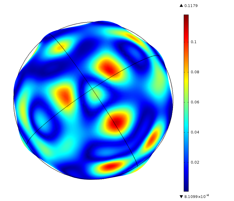
The modelled microbubble will vibrate in response to an applied field, rapidly expanding and contracting. The behaviour of the microbubble can be analysed as shell displacement, as shown in µm in the image below.
In addition to this, other scenarios and interactions can be modelled. For example, the image below shows a microbubble of 3 µm radius, located at a distance 0.6 µm away from a cell membrane.
A series of images show the effect the applied ultrasound signal has on the microbubble and, most importantly, its encapsulating shell in high and low pressure acoustic fields.
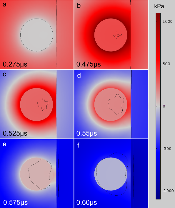
It can be seen from the image above, that in frames a and b (positive acoustic pressure regions) the microbubble contracts, and the microbubble and cell membrane move towards each other. This causes the encapsulating shell to buckle (b).
During the negative pressure phase (c, d, e, and f) the microbubble starts to expand and change shape. With the shell now fully buckled, the microbubble moves towards the cell membrane in a process similar to previously reported jetting behaviour.
The force applied to the cell membrane as a result of this can cause cell damage, or semi-permanant pores or holes in its structure. This is known as 'sonoporation' and can be used to aid uptake of drugs within specific targeted cells.
The use of acoustic waves and UCAs to cause pores in cell-membranes is often termed 'sonoporation'.
A major part of our research is primarily concerned with studying the mechanisms and parameters associated with therapeutic drug delivery using sonoporation. This not only includes simulating microbubble UCA behaviour, but also researching aspects of UCA construction, characterising the acoustic properties of UCA microbubbles, and imaging of UCA microbubble processes.
More information can be found in
G. Y. V. Léauté, S. Harput, J. McLaughlan, D. M. J. Cowell, S. Freear. A three-dimensional viscoelastic finite element model for microbubble non-spherical dynamics near a soft membrane. IEEE International Ultrasonics Symposium (IUS), 2012. In Press
See also our list of publications
read more...