Biomedical Imaging Example: Teeth
This research proposes an ultrasound contact imaging method, used to measure the enamel thickness of a human tooth.
The method demonstrates novel application of linear frequency modulated (LFM) 'chirp' signals, and the fractional Fourier transform (FrFT) - a signal processing technique that can be used to separate signals that are overlapping in time and frequency.
The aim of the work is to measure the thickness of the enamel layer, locate discontinuities, and produce a full image of a tooth ex-vivo using ultrasound imaging.
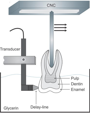
The figure shows a human tooth positioned on a computer numerically controlled (CNC) mechanical stage. A 15 MHz delay-line transducer was submerged within glycerin and positioned so as to contact the enamel of the tooth.
LFM chirp signals were chosen in this study to increase both the signal-to-noise ratio of the reflections and also penetration depth, therefore improving image quality.
Excitation signals were applied to the transducer using an arbitrary waveform generator and linear power amplifier. Reflections from the internal layers of the tooth were received in a pulse-echo mode.
Received signals were processed using matched filtering techniques and also the FrFT.
Matched filtering detects the coded LFM signal in the presence of other signals or noise
Processing with the FrFT allows separation of long-duration LFM chirp signals overlapping in time and frequency. Overlapping echoes are present due to successive reflections within the enamel and dentin layers.
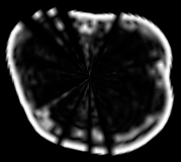
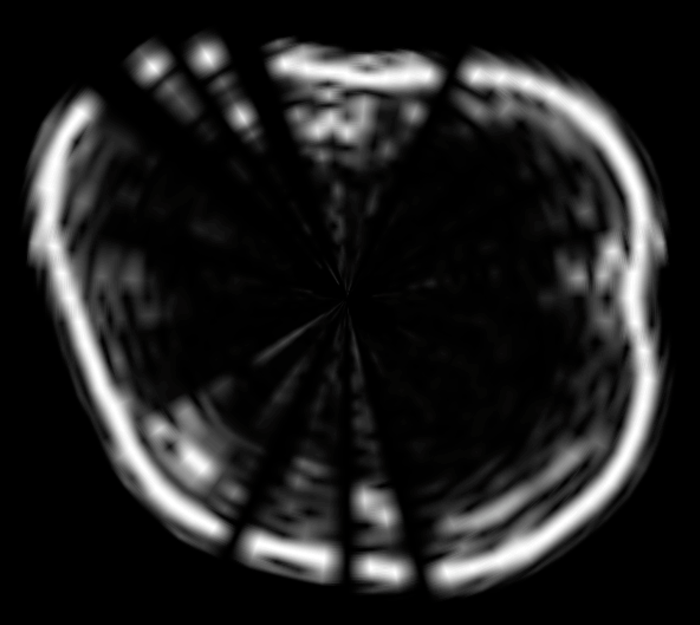
Images of the tooth processed using matched filtering only (left), and with both matched filtering and the FrFT (right) are shown below.
The Fourier transform transforms from time to frequency.
The FrFT is a modified version of the Fourier transform and can be used to transform to any line of angle in the time-frequency space.
Tooth image using matched filter processing.
Tooth image using matched filter and FrFT processing.
Results show that using matched filtering in combination with the FrFt produces more detailed images, and enables more accurate estimation of enamel thickness.
More information can be found in
S. Harput, Y. Evans, N. Bubb, S. Freear, Diagnostic ultrasound tooth imaging using fractional fourier transform, IEEE Transactions on Ultrasonics, Ferroelectrics and Frequency Control, Vol. 58, Issue: 10, pp. 2096 - 2106 DOI White Rose
S. Harput, T. Evans, N. Bubb, S. Freear, Detection of restoration faults under fillings in human tooth using ultrasound, IEEE International Ultrasonics Symposium (IUS) pp.1443 - 1446, 2011, DOI White Rose
See also our list of publications
read more...


